Clinical information
Bullous autoimmune dermatoses are rare, blister-forming diseases of the outer skin and the adjacent mucous membranes. They are characterised by the formation of autoantibodies against structural proteins of the skin. These structural proteins are responsible for the cell-to-cell contact of keratinocytes within the epidermis and the adhesion of the epidermis to the dermis. Bullous autoimmune dermatoses are divided into four main groups based on their target antigens and the localisation of the blisters:
- Pemphigus diseases: desmoglein 1 (Dsg1), desmoglein 3 (Dsg3), different plakins (esp. envoplakin)
- Pemphigoid diseases: BP180, BP230, laminin 332, p200
- Epidermolysis bullosa acquisita (EBA): collagen type VII
- Dermatitis herpetiformis: endomysium (tissue/epidermal transglutaminase), deamidated gliadin peptides (GAF-3X)
Diagnostics
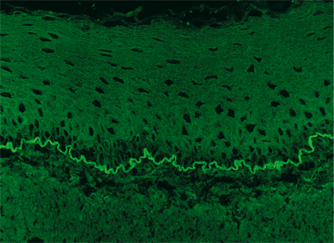 A conclusive diagnosis of blister-forming autoimmune dermatoses requires both the histopathological detection of tissue-bound autoantibodies by direct immunofluorescence and the serological determination of circulating autoantibodies. The disease-specific autoantibodies against epidermal antigens (prickle cell desmosomes and epidermal basement membrane) are detected using the indirect immunofluorescence test (IIFT) with tissue sections of primate oesophagus. For further differentiation of autoantibodies against basement membrane structures, tissue sections of primate salt-split skin are used. Final diagnosis is based on a combination of the clinical picture with the detection of autoantibodies against the specific target antigens using IIFT, monospecific ELISA or immunoblot analyses.
Patients who suffer from bullous pemphigoid (BP) exhibit autoantibodies against BP180 and frequently also against BP230. The serum level of autoantibodies against BP180 correlates with the disease activity of BP, while the serum level of autoantibodies against BP230 is associated with the duration of the disease. Autoantibodies against desmoglein 1 and 3 are markers for pemphigus diseases. IIFT has proven valuable for detecting circulating autoantibodies in pemphigus. ELISA offers the same sensitivity and specificity as IIFT if recombinant desmoglein 1 and 3 are used. The anti-Dsg1 and -Dsg3 antibody levels measured correlate to a large extend with the severity and activity of the disease and the therapy success. The determination of autoantibodies against envoplakin contributes to diagnosis of paraneoplastic pemphigus (PNP) as well as supports differential diagnostics. The detection of autoantibodies against collagen type VII confirms the diagnosis of EBA and enables differentiation from other bullous autoimmune dermatoses. Laminin 332 is the target antigen of a clinically relevant subform of mucous membrane pemphigoid. For differential diagnosis, anti-laminin 332 antibodies must be determined in suspected cases, which is made possible using the cell-based immunofluorescence test from EUROIMMUN.
Files
Autoimmune bullous dermatoses
Serological diagnosis of bullous autoimmune dermatoses
Diagnosis of mucous membrane pemphigoid


