Clinical information
Paraneoplastic neurological syndromes (PNS) are diseases of the central and peripheral nervous system that occur in direct association with tumours. However, they are not directly caused by the tumour or by its metastases, nor are they a side -effect of treatment with cytostatic drugs or radiotherapy. Approx. 15 % of patients with malignant diseases, especially lung tumours, have PNS.
Depending on the type of tumour, tumour cells express antigens such as amphiphysin, CV2 / CRMP5, PNMA2 (Ma2 / Ta), Ri, Yo, Hu, ZIC4 or Tr (DNER), which can induce the formation of specific autoantibodies. There is no known direct pathogenic effect of these autoantibodies. Instead, a T-cell-mediated immune response is involved in the neuronal degeneration. This also results in patients responding poorly to B-cell-based immunotherapy.
Diagnostics
New diagnostic criteria for PNS have recently been established (Graus et al., 2020). What is new is the subdivision into high-risk phenotypes (encephalomyelitis, limbic encephalitis, rapidly progressive cerebellar syndrome, opsoclonus myoclonus syndrome, sensory neuronopathy, gastrointestinal pseudoobstruction (enteric neuropathy), Lambert–Eaton myasthenic syndrome) and intermediate-risk phenotypes for PNS (including encephalitis, anti-NMDA receptor encephalitis, brainstem encephalitis, Morvan syndrome, isolated myelopathy, stiff-person syndrome, polyradiculoneuropathy).
The term “onconeural antibodies” has been replaced by “high-risk antibodies” for cancer (tumour in >70 % of patients with these antibodies) and “intermediate-risk antibodies” for cancer (tumour in 30 – 70 % of patients with these antibodies). The guidelines classify three levels of evidence for PNS: “definite”, “probable” and “possible”. Each level can be identified using the PNS-care score, which assesses the clinical phenotype, the antibody type, the presence or absence of cancer and the follow-up time. With the exception of opsoclonus myoclonus syndrome, the diagnosis of “definite PNS” requires the presence of high- or intermediate-risk antibodies.
Neuronal antibodies have been established as useful biomarkers and their detection is of paramount importance in the diagnosis of PNS. For a reliable diagnosis, the guidelines recommend that autoantibodies in PNS should always be determined using two independent methods.
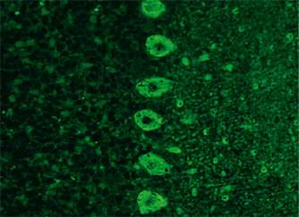
Gold standard methods include indirect immunofluorescence (IFA or immunohistochemistry) based on brain sections, for example, using specific BIOCHIP Mosaics from EUROIMMUN for neurology. For confirmation, immunoblots (e. g. EUROLINE profiles) for the monospecific detection of autoantibodies, mainly against intracellular antigens, or cell-based assays (CBAs) for the detection of antibodies against cell surface or synaptic proteins are used. Neuronal autoantibodies should be tested in both serum and cerebrospinal fluid.